Healthtech
From X-rays to CT scans: A Comprehensive Guide on How Radiologic Technology Works
Radiologic technology has come a long way since the discovery of X-rays over 100 years ago. Today, radiology plays an essential role in modern medicine, providing healthcare professionals with crucial insights into the inner workings of our bodies. From broken bones to cancerous growths, radiologic imaging can help diagnose and treat a wide range of conditions. But how does it work? In this comprehensive guide, we’ll take you through the science behind radiologic technology – from early X-ray machines to advanced CT scans.
Introduction to Radiologic Technology
Radiologic technology is the branch of medicine that uses radiation to diagnose and treat disease. The two main types of radiologic technologists are diagnostic medical sonographers and therapeutic radiographers. Diagnostic medical sonographers use ultrasound to create images of the body, while therapeutic radiographers use x-rays and other forms of radiation to treat cancer and other diseases.
Radiologic technologists must be licensed in most states. In order to become licensed, radiologic technologists must complete an accredited training program and pass a national exam. Some states also require licensure by the American Registry of Radiologic Technicians (ARRT). The ARRT offers certification in both diagnostic imaging and radiation therapy.
Once licensed, radiologic technologists can work in a variety of settings, including hospitals, clinics, private practices, and research facilities. They may also choose to specialize in a particular area of medicine, such as cardiology or pediatrics.
History of Radiologic Technology
The history of radiologic technology is a long and fascinating one. It’s a story of discovery, innovation, and adaptation that continues to this day.
Radiologic technology is a field of medicine that uses imaging to diagnose and treat diseases. It dates back to the early days of X-rays, when German physicist Wilhelm Röntgen discovered their existence in 1895.
Radiologic technology has come a long way since then. Today, there are many different types of imaging modalities that radiologists can use to visualize the human body, including X-rays, computed tomography (CT), magnetic resonance imaging (MRI), and ultrasound.
Each of these modalities has its own strengths and weaknesses, but all of them play an important role in diagnosing and treating a variety of conditions. In recent years, radiologic technologists have also begun to use artificial intelligence (AI) to help interpret images and make diagnoses.
Types of Imaging Tests Used in Radiology
There are many different types of imaging tests used in radiology, each with its own advantages and disadvantages. The most common types of imaging tests are X-rays, CT scans, MRIs, and ultrasounds.
X-rays are the most common type of imaging test. They are quick and easy to perform, and can be used to image many different parts of the body. However, X-rays cannot provide as much detail as other types of imaging tests.
CT scans are more detailed than X-rays, and can be used to image both soft tissue and bone. CT scans take longer to perform than X-rays, and may require the use of contrast dye.
MRIs provide even more detail than CT scans, and can be used to image both soft tissue and bone. MRIs take longer to perform than CT scans, and may require the use of contrast dye.
Ultrasounds are used to image soft tissue only. Ultrasounds do not use ionizing radiation, so they are considered safe for pregnant women and children. However, ultrasounds cannot provide as much detail as other types of imaging tests.
How Does an X-Ray Work?
X-rays are a type of electromagnetic radiation, just like visible light. They are produced when high-energy electrons hit a metal target inside an X-ray tube. The X-rays that are produced travel through the patient’s body and are absorbed to varying degrees by different tissues.
Some of the X-rays pass straight through the body and are detected by an X-ray film or digital detector on the other side. The darker areas on the film or digital image represent the parts of the body where more X-rays were absorbed. This produces an image of the inside of the body that can be interpreted by a radiologist.

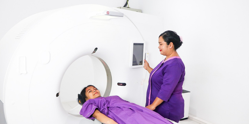
What Is a CT Scan and How Does It Work?
A CT scan, also known as a CAT scan, is a medical imaging procedure that uses X-rays and computer technology to create cross-sectional images of the body. CT scans are often used to diagnose problems with the bones, organs, and blood vessels.
How does a CT scan work? A CT scanner consists of a large doughnut-shaped machine that surrounds the patient’s table. The table moves slowly through the machine while it rotates around the patient, taking multiple X-ray images. These images are then sent to a computer where they are reconstructed into cross-sectional images of the body.
CT scans are painless and typically take 30-60 minutes to complete. During the procedure, patients may be asked to hold their breath for short periods of time to avoid blurring the images.
Benefits of Radiologic Technology
Radiologic technologists are highly trained professionals who use these imaging modalities to help physicians diagnose and treat their patients. Radiologic technology offers many benefits, both for patients and healthcare providers.
Patients benefit from radiologic technology because it allows for early detection of many medical conditions. Imaging can often detect problems before they cause symptoms, which means that treatment can be started sooner. In some cases, imaging can even be used to prevent problems from developing in the first place. For example, mammograms can detect breast cancer at an early stage, when it is most treatable.
Radiologic technology also offers many benefits for healthcare providers. Imaging allows doctors to get a detailed look at a patient’s anatomy without having to perform surgery. This means that diagnosis and treatment can be completed more quickly and with less invasiveness. In addition, imaging provides doctors with valuable information that can be used to plan surgeries and other procedures.
Potential Risks Associated With Radiologic Technology
There is always a potential for risks when undergoing any type of medical procedure, and radiologic technology is no exception. While the risks are generally low, there is a small possibility of developing cancer from exposure to ionizing radiation. Other potential risks include skin reactions, pregnancy complications, and organ damage.
It’s important to discuss any potential risks with your doctor prior to having a radiologic procedure performed. This will help you make an informed decision about whether or not the procedure is right for you.
Tips for Preparing for a Radiology Test
If you’re scheduled for a radiology test, there are a few things you can do to prepare. First, it’s important to understand what the test is and why your doctor has ordered it. This will help you know what to expect. You should also ask your doctor or the technologist if there are any special instructions you need to follow before the test. For example, you may need to fast for a certain period of time before having a contrast CT scan.
Once you know what the test involves, you can take steps to make sure it goes smoothly. For example, if you’re claustrophobic, let your doctor or technologist know so they can take measures to help you feel more comfortable during the scan. If you’re having an X-ray, you may be asked to remove any jewelry or clothing that contains metal. And if you’re pregnant or think there’s a possibility you could be pregnant, be sure to tell your doctor or technologist so they can tailor the exam accordingly.
By following these tips and working with your medical team, you can ensure that your radiology test goes as smoothly as possible.
Conclusion
Radiologic technology is an invaluable tool in the medical world, helping doctors diagnose and treat a range of ailments. This comprehensive guide has explored how X-rays work, what CT scans are used for and their differences. We hope this article has given you a better understanding of radiology techniques so that you can make informed decisions about your healthcare needs. With advances in radiologic technology happening all the time, there’s sure to be even more effective treatments available in the future – making it easier than ever to stay healthy and get back on track with your life!


Stellar Repair for QuickBooks – An Effective Software to Repair Corrupted QBW Files

The Ultimate Dubai Watch Hunt: How to Score the Best Deals on Used Luxury Timepieces

Architectural Wonders: The Work of Hampshire’s Finest Architects

The Dynamics of CryptoGrab: How It Works

How to Choose the Right Aged Care Facility for Your Loved Ones
Elevate Your Image With Corporate Brand Photography

Celebrating Vision and Innovation: The 2nd Annual 4BIDDEN Conscious Awards

What to Expect When Visiting an Endodontist Los Angeles

What Are the Cons of Creating Your Own Website?

How Hiring a Fridge Can Benefit Your Startup

Breaking News: Private Helicopter Tours Service In Atlanta Is Fusing With M.V.P Atlanta | Dujaun Hayles Is Elevating Atlanta’s Luxury Vehicle Experience

Site GPT: Redefining Website Content Creation with AI

Importance of Digital Marketing for Small and Medium Enterprises (SMEs)

Everything You Need To Know About Enterprise Resource Planning Software

The Market Heading Towards Mass Institutionalization of the Crypto Industry, Says Ape Terminal Founder Hassan Hatu Sheikh

Exploring the Stability and Advantages of USDT in Financial Management

Protecting Your Data – Insights from the Vans Cybersecurity Breach

The Puzzling Story Behind Ezras Lightsaber: Divulging the Secrets of This Famous Weapon

Top Dog Marketing Online Mistakes to Avoid for a Successful Business
