Introduction
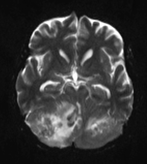
Magnetic Resonance Imaging (MRI) has been a cornerstone in the field of medical imaging, providing detailed anatomical information for the diagnosis and management of various conditions. In recent years, a specialized MRI technique called Diffusion-Weighted Imaging (DWI) has gained prominence in the assessment of ankle tumors. DWI MRI offers unique insights into tissue microstructure and cellularity, allowing for early and accurate detection, characterization, and monitoring of ankle tumors. This article explores the significance of DWI-MRI in ankle tumor imaging and its potential to revolutionize the way we approach these conditions.
Ankle Tumors: A Diagnostic Challenge
Ankle tumors can encompass a broad spectrum of pathologies, ranging from benign cysts and lipomas to malignant lesions such as sarcomas or metastatic disease. Accurate diagnosis and characterization of ankle tumors are essential for treatment planning and patient outcomes. However, conventional MRI techniques often have limitations when it comes to distinguishing between different tumor types and assessing their aggressiveness.
The Role of DWI-MRI
DWI-MRI is a specialized imaging technique that provides unique information about the movement of water molecules within tissues. In the context of ankle tumor imaging, this technique can be a game-changer. Here’s why:
Tissue Microstructure: DWI-MRI is highly sensitive to changes in tissue microstructure. It can detect alterations in cell density, cellular integrity, and tissue architecture, making it a valuable tool for distinguishing between benign and malignant ankle tumors.
Early Detection: DWI-MRI can detect subtle changes in tissue before they become apparent on conventional MRI sequences, allowing for earlier diagnosis and intervention.
Differentiation of Tumor Types: Different tumor types have distinct DWI-MRI characteristics. For example, benign tumors typically exhibit low DWI signal, while malignant tumors tend to show high signal intensity. This differentiation is crucial for treatment planning.
Assessment of Treatment Response: DWI-MRI can also be used to monitor the response to therapy. Changes in tumor cellularity and vascularity can be assessed over time, helping clinicians adapt treatment strategies as needed.
Clinical Applications
The clinical applications of DWI-MRI in ankle tumor imaging are manifold:
Preoperative Planning: Surgeons can benefit from DWI-MRI by gaining a better understanding of tumor margins and the extent of infiltration into surrounding tissues, which is crucial for surgical planning.
Tumor Characterization: DWI-MRI aids in characterizing tumors as benign or malignant, helping to guide treatment decisions. For instance, a benign cyst can be confidently differentiated from a malignant soft tissue sarcoma.
Monitoring Disease Progression: For patients undergoing non-surgical therapies like chemotherapy or radiation, DWI-MRI allows for real-time monitoring of tumor response and adaptation of treatment strategies based on the observed changes.
Surveillance: In cases where ankle tumors are known to recur, regular DWI-MRI scans can help detect recurrences at an early stage, improving long-term outcomes.
Challenges and Considerations
While DWI MRI holds immense promise in ankle tumor imaging, there are some challenges to consider:
Technical Expertise: Performing and interpreting DWI-MRI requires specialized training and expertise, which may not be available at all medical facilities.
Artifacts: DWI-MRI can be sensitive to motion artifacts and may require longer scanning times, potentially causing discomfort for the patient.
Cost: Advanced imaging techniques like DWI-MRI can be more expensive than conventional MRI, which may limit access for some patients.
Conclusion
Diffusion-Weighted Imaging (DWI-MRI) is emerging as a valuable tool in the field of ankle tumor imaging. Its ability to provide insights into tissue microstructure and cellularity makes it a powerful diagnostic and monitoring tool. As technology continues to advance and become more accessible, DWI-MRI is likely to play an increasingly significant role in the early detection, characterization, and treatment of ankle tumors. This promises improved patient outcomes and a better understanding of these complex conditions.


