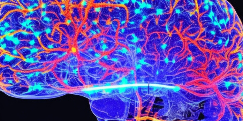
Are you curious about how scientists gain insight into the inner workings of the human brain? Wonder no more! Neuroimaging techniques have revolutionized our understanding of the brain, enabling us to visualize its activity in real-time. These methods have opened up new possibilities for research and the treatment of neurological disorders. In this article, we will explore the most common neuroimaging techniques used today, their advantages, and their limitations.
Introduction:
Neuroimaging techniques have revolutionized our understanding of the brain by allowing us to visualize its activity in real-time. These advanced methods have opened up new avenues for research and treatment of neurological disorders. From functional magnetic resonance imaging (fMRI) to positron emission tomography (PET) and single-photon emission computed tomography (SPECT), we will uncover the strengths and limitations of each method. Join us as we embark on a journey to understand the power of neuroimaging in unraveling the mysteries of the human brain.
Neuroimaging Techniques: An Overview
Neuroimaging techniques encompass a range of non-invasive tools that allow scientists to study and visualize the human brain. With these methods, researchers can examine brain structure, function, and connectivity in real-time. There are several types of neuroimaging techniques commonly used today, each with its own unique strengths and weaknesses.
Functional Magnetic Resonance Imaging (fMRI) is a technique that measures changes in blood flow within the brain, providing insights into neural activity during cognitive processes like attention and memory recall.
Positron Emission Tomography (PET) uses radioactive tracers to map metabolic activity in specific brain regions, aiding in the diagnosis of conditions such as Alzheimer’s disease.
Single-Photon Emission Computed Tomography (SPECT) also utilizes tracers to visualize blood flow and metabolic activity, often used alongside other imaging techniques for a comprehensive understanding of brain function.
Magnetoencephalography (MEG) measures the magnetic fields generated by electrical currents in neurons, offering high temporal resolution but relatively lower spatial resolution compared to other methods.
Each neuroimaging technique possesses distinct advantages and disadvantages, depending on the specific aspects of brain function under investigation.
Functional Magnetic Resonance Imaging (fMRI)
Functional Magnetic Resonance Imaging (fMRI) is a non-invasive technique that detects changes in blood flow to measure brain activity. This revolutionary method has significantly advanced cognitive neuroscience by enabling researchers to explore the involvement of different brain regions in complex mental processes such as decision making and language comprehension.
During an fMRI scan, a strong magnetic field aligns hydrogen atoms in the body. By applying radio waves to perturb these atoms, they emit detectable energy. By tracking changes in this signal over time, scientists can infer which areas of the brain are active during specific tasks or stimuli.
One advantage of fMRI is its high spatial resolution, capable of detecting activity at a resolution of a few millimeters. However, it comes with a lower temporal resolution compared to techniques like electroencephalography (EEG). Additionally, fMRI requires participants to remain still inside a loud and sometimes claustrophobic scanner for extended periods.
Despite these limitations, fMRI remains one of the most powerful tools available for studying human cognition and behavior.
Positron Emission Tomography (PET)
Positron Emission Tomography (PET) is a neuroimaging technique that utilizes radioactive tracers to visualize brain activity. By detecting the gamma rays emitted during the decay of positrons in brain tissue, PET scans provide valuable insights into brain function.
During a PET scan, a small amount of radioactive material is injected into the patient’s bloodstream, binding to receptors or specific molecules associated with brain functions of interest. Images are then captured while the patient is at rest or performing tasks.
PET offers the ability to measure changes in neurotransmitter function, providing valuable information about how different compounds affect brain activity. Additionally, it can detect early signs of disease before structural damage occurs.
However, PET has limitations, including its high cost and limited availability at some medical facilities. Additionally, radiation exposure during scans may restrict its use for repeated studies or young patients.
Despite these drawbacks, Positron Emission Tomography remains an essential tool for understanding and diagnosing neurological disorders.
Single-Photon Emission Computed Tomography (SPECT)
Single-Photon Emission Computed Tomography (SPECT) is a neuroimaging technique that employs radioactive tracers to create 3D brain images. By detecting gamma rays emitted during tracer decay, SPECT enables visualization of blood flow and metabolic activity.
SPECT’s advantage lies in its ability to detect changes in brain function over time, making it useful for studying progressive disorders like Alzheimer’s disease. It can also identify abnormalities in blood flow that may indicate the presence of tumors or other lesions.
However, SPECT has limitations. Its resolution is lower compared to newer techniques like fMRI, providing less detailed information about specific brain regions. Additionally, repeated use of SPECT raises concerns due to radiation exposure.
Nevertheless, SPECT remains a valuable tool for neuroscience research and clinical diagnosis, shedding light on how different brain areas function under various conditions or during specific tasks.
Magnetoencephalography (MEG)
Magnetoencephalography (MEG) is a neuroimaging technique that measures the magnetic fields generated by neuronal activity in the brain. Unlike other techniques that rely on changes in blood flow or glucose metabolism, MEG directly captures the electrical activity of neurons.
MEG offers several advantages, including high temporal resolution, allowing researchers to observe rapid changes in neural activity. It is safe for repeated use as it does not involve ionizing radiation.
However, MEG has limitations. It can only detect activity from cortical areas near the brain’s surface due to magnetic field attenuation. Additionally, while MEG provides excellent spatial resolution when combined with MRI or CT scans, it cannot provide structural information on its own.
Magnetoencephalography is an incredibly powerful tool for understanding how different brain regions communicate and interact at a fine level of detail.
How Do These Techniques Work?
Neuroimaging techniques are vital for understanding the intricate functions of the brain. But how exactly do these methods operate? Let’s explore the principles behind them.
Functional Magnetic Resonance Imaging (fMRI) is based on the principle that when neurons in a specific brain region become active, blood flow to that area increases. By measuring changes in blood oxygen levels, fMRI indirectly indicates neural activity.
Positron Emission Tomography (PET) involves injecting radioactive tracers into the bloodstream. These tracers bind to glucose molecules and emit positrons, which sensors outside the brain detect. By tracking the origin of these signals, researchers generate images of brain activity.
Single-Photon Emission Computed Tomography (SPECT) follows principles similar to PET but uses different tracers. It detects changes in cerebral blood flow and metabolism, providing valuable information about neurological disorders.
Magnetoencephalography (MEG) measures the magnetic fields generated by electrical currents in neurons using highly sensitive sensors. This non-invasive method offers information about neuronal activation patterns with high temporal resolution.
Each neuroimaging technique has its strengths and limitations, making them suitable for different types of data collection in experiments or clinical investigations.
Advantages and Disadvantages of Each Technique
Functional Magnetic Resonance Imaging (fMRI) is a non-invasive technique that produces high-resolution real-time images of the brain. It excels in showing changes in blood flow, indicating increased neural activity. However, it is susceptible to motion artifacts and can produce noisy data.
Positron Emission Tomography (PET) involves injecting a radioactive tracer into the bloodstream to track brain metabolic activity. One advantage is its ability to detect low levels of activity, but it comes with drawbacks like radiation exposure and limited spatial resolution due to scanner size.
Single-Photon Emission Computed Tomography (SPECT) functions similarly to PET but employs different tracers and allows longer scanning periods. It has lower radiation exposure than PET but offers poorer spatial resolution.
Magnetoencephalography (MEG) measures the magnetic fields generated by electrical currents in neurons. Its main advantage is its high temporal resolution, enabling the observation of rapid changes in neural activity. However, MEG measurements are vulnerable to interference from environmental factors and necessitate specialized equipment.
Each neuroimaging technique possesses unique strengths and weaknesses, rendering them suitable for specific research questions or clinical applications based on prioritized aspects.
Conclusion
Neuroimaging techniques have revolutionized our understanding of the brain and its functions. These methods enable visualization of different brain regions’ activity, providing valuable insights into the neural processes underlying various cognitive and behavioral functions.
Each technique has its own advantages and disadvantages, making them suitable for different types of studies. For instance, fMRI is ideal for investigating changes in blood flow during specific tasks or stimuli, while PET can detect changes in neurotransmitter levels associated with diseases or disorders.
As technology advances, we can expect even more sophisticated neuroimaging techniques to emerge, providing us with a deeper understanding of the brain’s workings. As a society, it is crucial to continue investing resources in this field to ensure future generations benefit from these discoveries.


