Medical imaging has come a long way in aiding healthcare professionals to diagnose and treat a wide range of conditions. Magnetic Resonance Imaging (MRI) is one such technology that has revolutionized the field of diagnostic medicine. Within the realm of MRI, there are different types and sequences used to capture images of the human body. Two of the most commonly used MRI sequences are T1 and T2 weighted imaging. In this blog post, we’ll explore the differences between T1 VS T2 MRI, how they work, and their respective clinical applications.
The Basics of MRI
Before delving into the differences between T1 and T2 MRI, let’s briefly understand how MRI works. MRI uses a strong magnetic field and radio waves to create detailed images of the internal structures of the body. The human body is composed mostly of water, and the hydrogen atoms in water have protons that behave like tiny magnets. When placed in a magnetic field and exposed to radio waves, these protons emit signals that can be captured and processed into detailed images.
T1 Weighted MRI
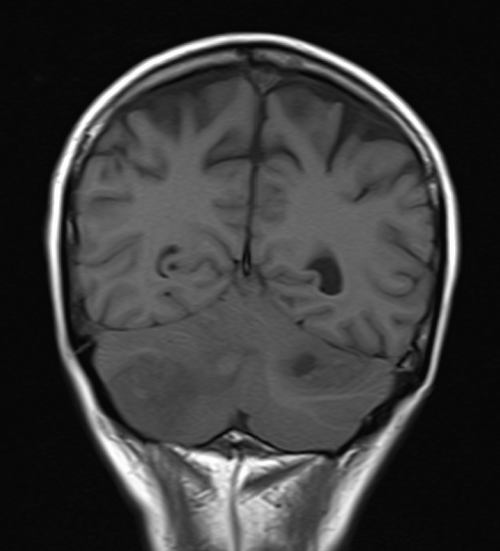
T1-weighted MRI, also known as T1 imaging or T1-weighted imaging, emphasizes the differences in the recovery times of hydrogen protons in different tissues. In T1 imaging:
- Fat appears bright: T1-weighted images are excellent at highlighting fat tissue, making it appear bright white.
- Fluid appears dark: Cerebrospinal fluid (CSF), which surrounds the brain and spinal cord, and other fluids, appear dark in T1-weighted images.
- Tissues vary in shades of gray: T1-weighted images differentiate various tissues based on their T1 relaxation times. Muscle, for example, appears intermediate in intensity.
Clinical Applications of T1-weighted MRI:
- Anatomical imaging: T1-weighted MRI is often used for high-resolution anatomical imaging, helping identify structures and lesions in the brain, spine, and musculoskeletal system.
- Evaluation of fatty tissues: T1-weighted images are essential in assessing fat content in organs, aiding in the diagnosis of conditions like fatty liver disease.
T2 Weighted MRI
T2-weighted MRI, on the other hand, focuses on the differences in the decay times of hydrogen protons in tissues. In T2 imaging:
- Fluids appear bright: Unlike T1 images, T2-weighted images make fluids, including CSF, appear bright.
- Fat appears dark: In T2-weighted images, fat appears dark or hypo-intense.
- Tissues vary in shades of gray: Similar to T1 images, T2-weighted images differentiate tissues based on their T2 relaxation times. Muscle, for instance, appears gray.
Clinical Applications of T2-weighted MRI:
- Detection of edema and inflammation: T2-weighted images are highly sensitive to edema (swelling) and inflammation, making them useful in identifying these conditions in various organs.
- Brain imaging: T2-weighted MRI is crucial for evaluating brain lesions, particularly in detecting multiple sclerosis (MS) plaques.
- Cardiac imaging: T2-weighted imaging can be used to assess myocardial edema in conditions like myocarditis.
Key Differences at a Glance
- Tissue contrast: T1-weighted MRI provides excellent contrast for fat and is suitable for anatomical imaging, while T2-weighted MRI is better for highlighting fluids and detecting edema and inflammation.
- Signal intensity: In T1-weighted images, fat is bright, and fluids are dark, whereas in T2-weighted images, it’s the opposite: fluids are bright, and fat is dark.
- Clinical applications: T1-weighted MRI is often used for structural assessments and evaluation of fat content, while T2-weighted MRI is preferred for detecting pathology, inflammation, and edema.
In summary, T1 VS T2 MRI sequences offer different insights into the human body, each with its unique clinical applications. Radiologists and healthcare professionals select the appropriate sequence based on the specific diagnostic information they need. Understanding the differences between these MRI types is crucial for accurate diagnosis and treatment planning in modern medicine.